Laboratory of X-Ray Methods
Head of the Laboratory, Dr. Alexander A. KARABTSOV
Tel: +7-(423) 231-71-32
E-mail: This email address is being protected from spambots. You need JavaScript enabled to view it.
The X-ray Laboratory provides high quality analytical work to support scientific activity of the scientists of FEGI. The main task of the laboratory is composition and structure of minerals, rocks, metals and their alloys investigation by methods of radiographic analysis, X-ray microanalysis, X-ray fluorescence analysis, and X-ray computer microtomography. At once the laboratory supports several projects including: (1) Gemstones of the Far East (mineralogy and formation conditions); (2) Experimental modeling of physical and chemical conditions critical to the formation of precious metal and other deposits. In addition, the laboratory cooperates closely with scientists of the Chemical Institute, Pacific Ocean Institute, and other institutes and subdivisions of the Far East Branch of RAS.
 |
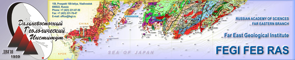
Desktop microtomograph Bruker SKYSCAN 1272 allows visualizing and measuring space structures of samples without their chemical and mechanical treatment. 256 gray gradations show virtual cross-section reconstruction of the object, based on the shaded (transmitted) images of a sample to be obtained during the analysis process.
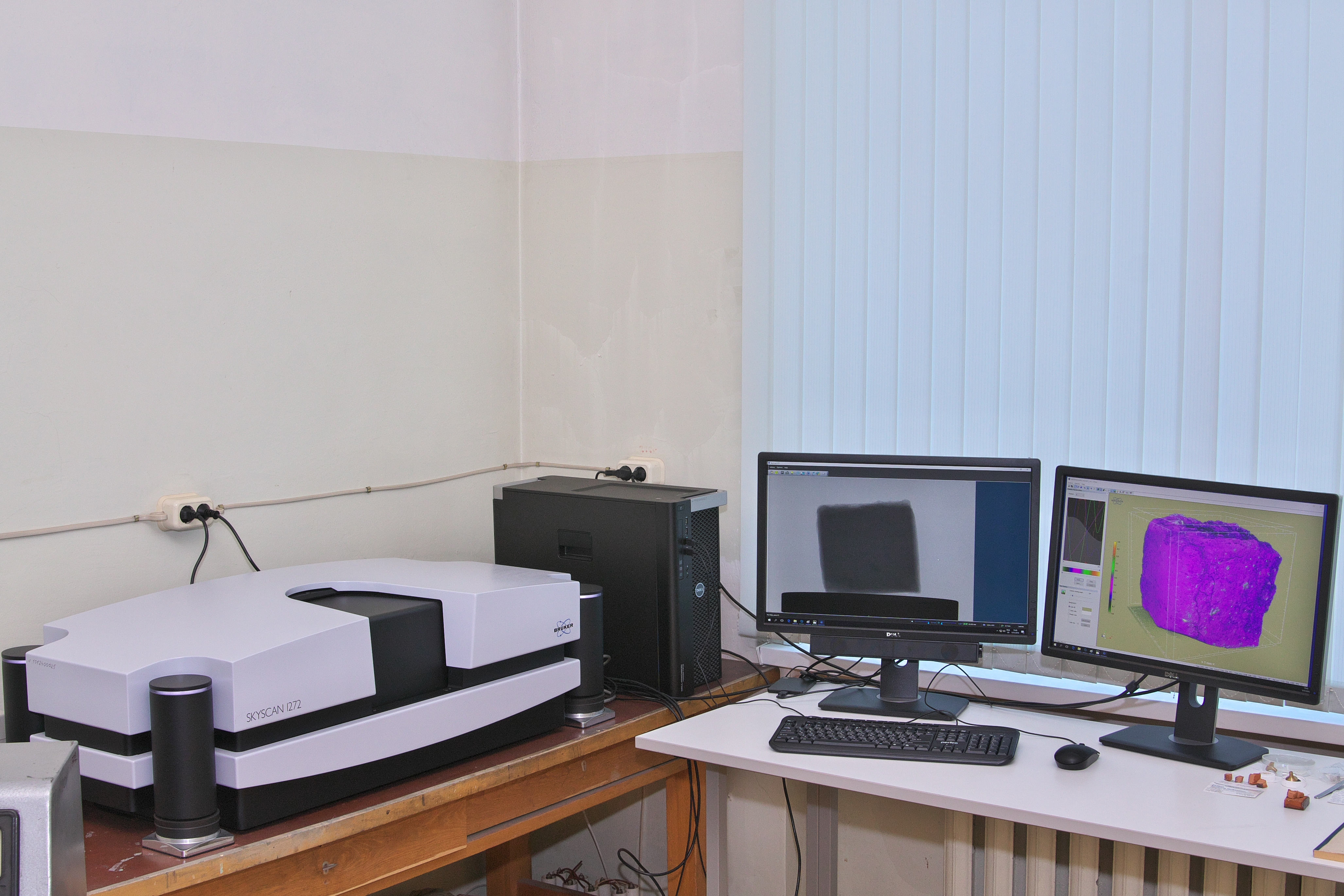
Electron probe X-ray microanalysis
Electron probe X-ray microanalysis (EPMA) is a non-destructive, local (5-10 micron) method for qualitative and quantitative analysis of element composition within a limited size at contents from 0.01 to 100 wt %. A very small amount of material is used for an electron microprobe measurement with the required sample size for complete analysis ranging from 10-13-10-16 g. Elements from B to U can be analyzed.
Both minerals and synthetic compounds can be analyzed, as well as micro inclusions from an uncovered surface of 1-5 microns in diameter.
The laboratory carries out electron probe X-ray microanalysis using a JEOL JXA-8100 electron microprobe with three wavelength dispersive spectrometers and the energy dispersive X-ray Spectrometer INCA-sight from Oxford Instruments (UK). The current spectrometer crystal inventory consists of: LIF, PET, TAP, and LDE2. The microanalyzer is also equipped with a cathodoluminescent Gatan attachment.
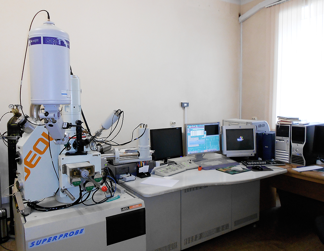 |
Radiographic analysis
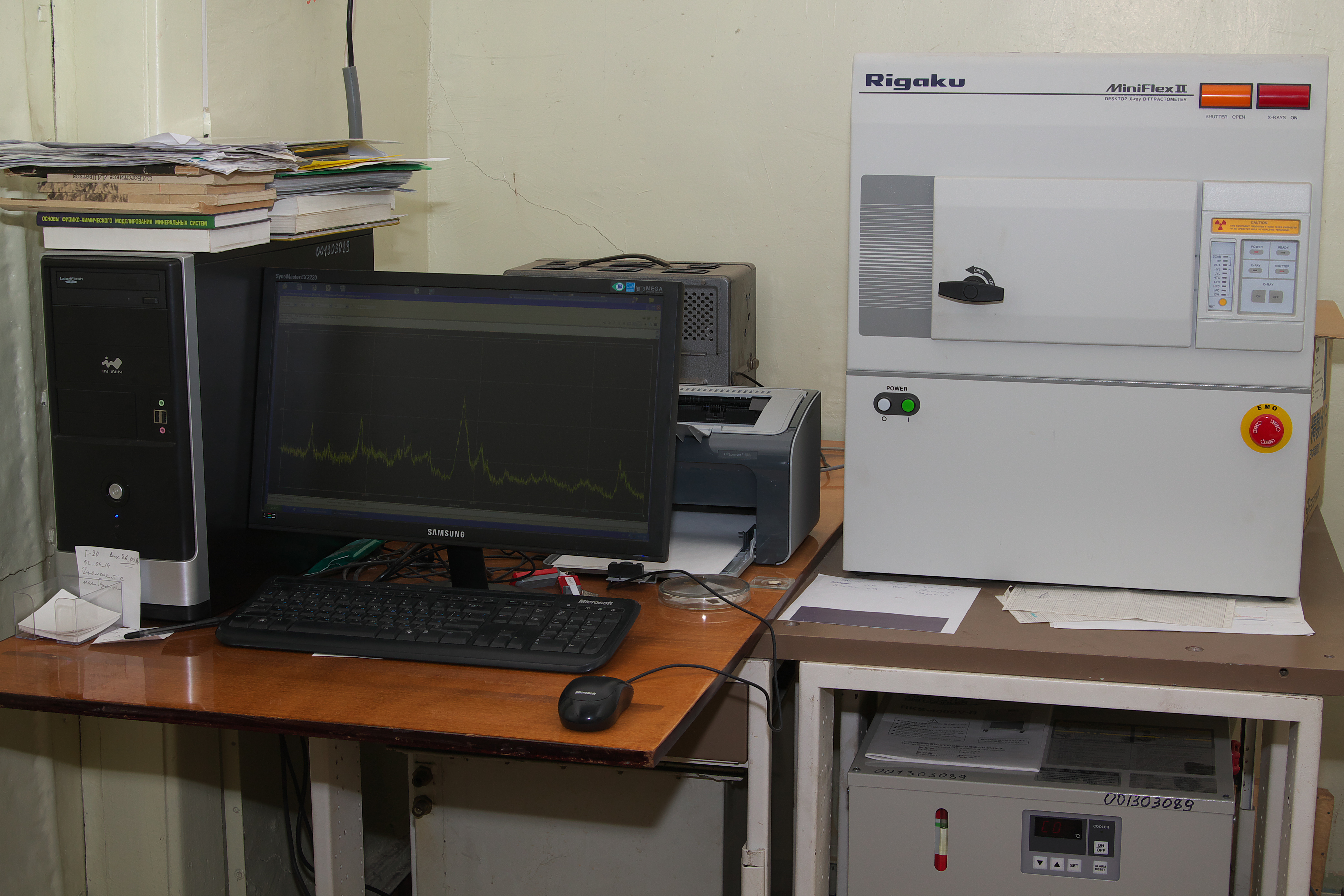
Radiographic analysis of polycrystals is one of the basic methods investigating composition and structure of solid substances, used in the laboratory. In many cases this method provides accurate and authoritative information in regard to the phase composition and structural features of a given material, which can’t be obtained using other analytical methods.
The laboratory carries out X-ray diffraction studies at the diffractometers DRON-3 (Russia) with monochromatic X-ray radiation, Rigaku Mini Flex II X-ray diffractometer, and Bruker D8-Discover (Germany).
 |
 |
X-Ray Fluorescence Analysis
X-ray fluorescence analysis (XRF) is a non-destructive physical method of qualitative and quantitative determination on the concentrations of micro- and macro elements in various rocks, minerals, ores and different-type mineral raw materials, soils, ashes, etc.. The method is based on irradiation of the investigated sample with electromagnetic radiation in the x-ray region of the spectrum and subsequent analysis of the spectrum of secondary X-ray radiation.
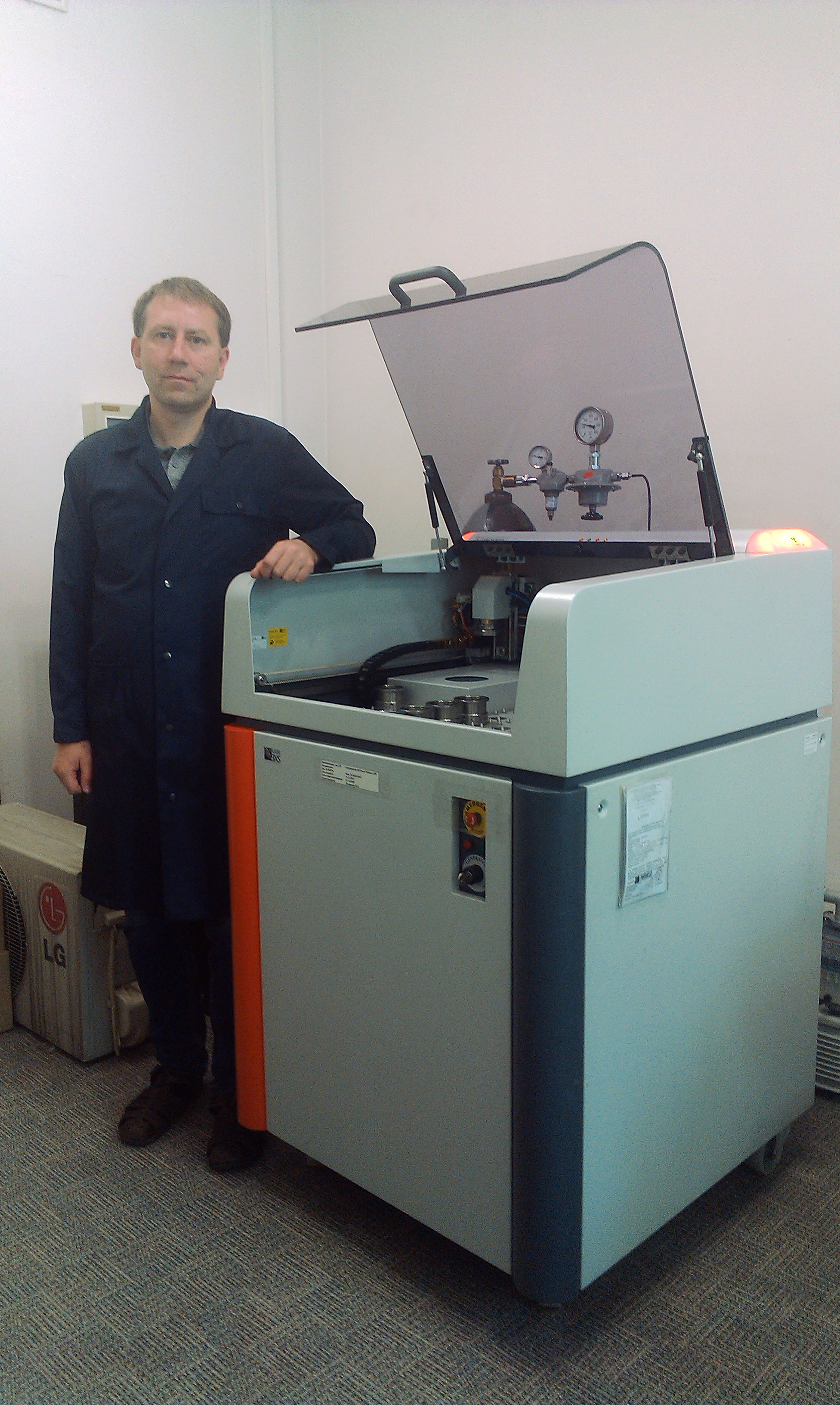 |
S4 Pioneer sequential X-ray spectrometer (Bruker AXS GmbH, Germany) is available for the X-ray fluorescence analysis. The device has an X-ray tube, capacity 4 kW, with Rh-anode and a thin (75 micron) beryllium window. The spectrometer has the following configuration: five analyzing crystals (LiF200, LiF220, Ge, PET, and OVO-55); two collimators with divergence to be 0.23 and 0.46 ; and eight primary X-ray filters: Cu (1000, 300, and 200 microns) and Al (800, 500, 200, 100, and 12.5 microns).
The laboratory performs quantitative determinations on the concentrations of petrogenetic elements Na, Mg, Al, Si, P, K, Ca, Ti, Mn, Fe and microelements S, Cl, V, Cr, Co, Ba, Rb, Sr, Y, Zr, Nb, As, Pb, Th, U, Ni, Cu, Zn, Ga in various rocks, minerals, ores and soils. For these purposes compressed tablets and fused disks are used. Detection limits of elements mostly are 2-10 ppm